
Craniofacial Development & Disease
Craniofacial anomalies are associated with approximately one-third of all birth defects. We use avian, murine and human induced pluripotent stem cell models to understand the cellular and molecular factors that contribute to facial development.
Image: (Top left) Control avian embryo, (Top right) talpid2 avian embryo with cleft lip and polydactyly. (Bottom left) wild-type, (middle) Ift88f/f;Crect, (right) Ift88f/f;Wnt1-Cre mouse embryos. Courtesy of Betsy Schock and Ching-Fang Chang.
Primary cilia & Ciliopathies
Primary cilium are ubiquitous, microtubule-based organelles used by all cells to integrate and transduce molecular signals. Our research program focuses on identifying how primary cilia function during facial development. Check out our database characterizing ciliary heterogeneity.
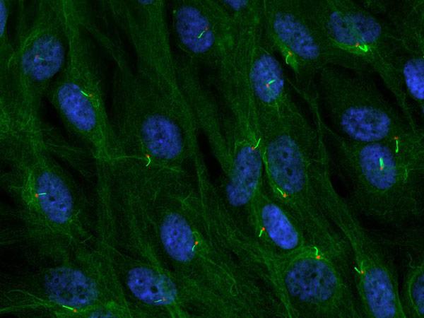
Image: Ciliary axoneme (green) and basal body (red) on neural crest stem cells. Courtesy of Ching-Fang Chang.
Neural Crest Stem Cells
Neural crest stem cells (NCSCs) are a transient population of cells that harbor stem cell properties, such as self-renewal and multipotency. We explore the molecular signals that guide NCSC differentiation into skeletal and neuronal derivatives.
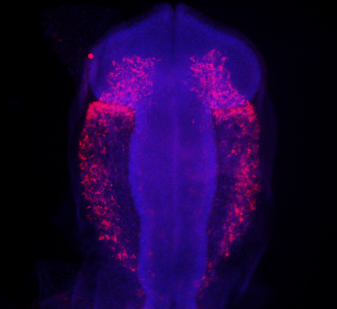
Image: Migrating neural crest stem cells (red) in an avian embryo. Courtesy of Betsy Schock.
Hedgehog signalling
The Hedgehog (Hh) signalling pathway has been intricately linked to the primary cilium and proper craniofacial development. We are interested in better understanding the Hh pathway in hopes of identifying mechanisms to alleviate craniofacial phenotypes.
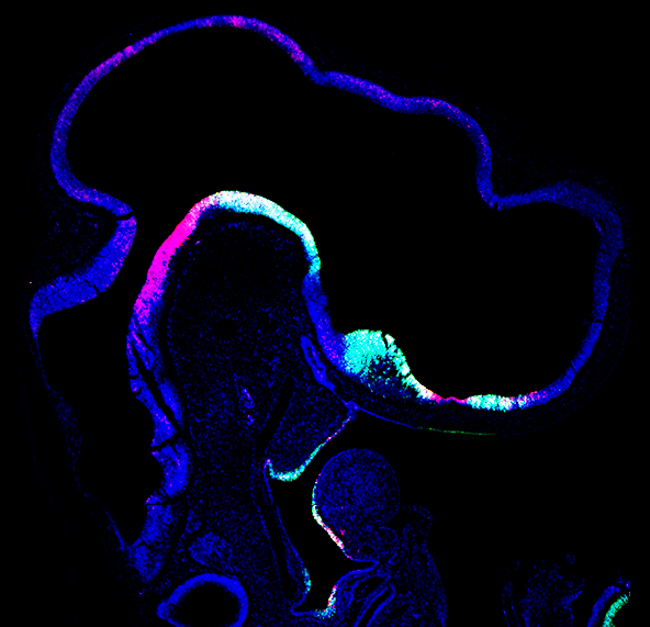
Image: Sagittal section of an avian embryo with Shh (green) and Ptch (red). Courtesy of Anne Carroll.